
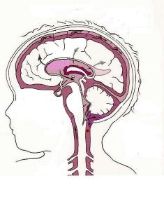
Hydrocephalus comes from the Greek: "hydro" means water, "cephalus" means head. Hydrocephalus is an abnormal accumulation of cerebrospinal fluid (CSF) within cavities called ventricles inside the brain. CSF is produced in the ventricles, circulates through the ventricular system and is absorbed into the bloodstream. CSF is in constant circulation and has many important functions. It surrounds the brain and spinal cord and acts as a protective cushion against injury. CSF contains nutrients and proteins necessary for the nourishment and normal function of the brain. It also carries waste products away from surrounding tissues. Hydrocephalus occurs when there is an imbalance between the amount of CSF that is produced and the rate at which it is absorbed. As the CSF builds up, it causes the ventricles to enlarge and the pressure inside the head to increase.
Hydrocephalus that is congenital (present at birth) is thought to be caused by a complex interaction of environmental and perhaps genetic factors. Aqueductal stenosis and spina bifida are two examples. Acquired hydrocephalus may result from intraventricular hemorrhage, meningitis, head trauma, tumors and cysts. Hydrocephalus is believed to occur in about 2 out of 1,000 births. The incidences of adult-onset hydrocephalus and acquired hydrocephalus are not known.
How Is Hydrocephalus Treated?
There is no known way to prevent or cure hydrocephalus. The most effective treatment is surgical insertion of a shunt. Learn more about shunts. Endoscopic third ventriculostomy (ETV) is growing in popularity as an alternative treatment method for hydrocephalus. Learn more about third ventriculostomy.
Hydrocephalus in Infants and Children
Hydrocephalus in infants and young children is frequently diagnosed at birth or shortly thereafter, but sometimes it is not diagnosed until the child is a little older.
With the advent of sophisticated imaging technologies such as magnetic resonance imaging (MRI) and ultrasonography, hydrocephalus can be diagnosed in utero, before the baby is born.
Signs and Symptoms of Hydrocephalus
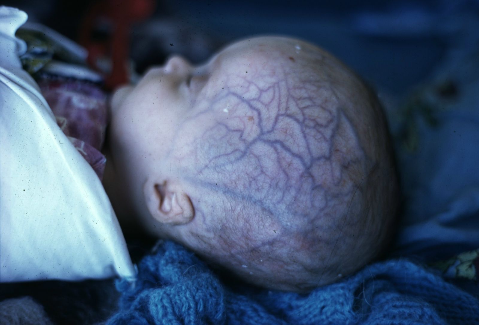
In an infant, the most obvious sign of hydrocephalus is an abnormal enlargement of the baby's head. The soft spot (fontanel) may be tense and bulging. The scalp may appear thin and glistening, and the scalp veins may appear to have unnatural fullness (prominence), as well. When you feel your baby's head along the suture lines, you may find that the bones are separated. Symptoms to watch for are vomiting, sleepiness, irritability and downward deviation of the baby's eyes (the sunsetting sign).
Toddlers whose sutures have not yet closed also show the signs of head enlargement. Older toddlers and children, once their sutures have closed, will show other symptoms of raised intracranial pressure (ICP) caused by their enlarged ventricles. Often these symptoms include headache, nausea, vomiting and sometimes blurred or double vision. The child might have problems with balance, delayed development in such areas as walking or talking, or poor coordination. As with infants, a child may be more irritable or tired than normal. The child may show a change in personality or be unable to concentrate or remember things, and their school performance may decline. Older children may have difficulty waking up and staying awake. While at times the symptoms are very noticeable, other times they can be very subtle and progress so slowly that only in retrospect are they appreciated.
Causes of Hydrocephalus
A variety of medical problems can cause hydrocephalus. In many children the problem is there at birth - this kind of hydrocephalus is referred to as congenital. Most cases of congenital hydrocephalus are thought to be caused by a complex interaction of genetic and environmental factors. Hydrocephalus that develops later in life in some children, and even in adults, but is caused by a condition that existed at birth, is still considered a form of congenital hydrocephalus. When hydrocephalus develops after birth and is caused by a factor such as head injury, meningitis or a brain tumor, it is termed acquired hydrocephalus. Parents must not blame themselves for their child's hydrocephalus. In almost all cases the circumstances contributing to a child's condition are beyond the parent's control.
Aqueductal Obstruction (Stenosis): The most common cause of congenital hydrocephalus is obstruction of the cerebral aqueduct - the long, narrow passageway between the third and fourth ventricle. Aqueductal obstruction may result from narrowing or blockage of the aqueduct, or may be caused by infection, hemorrhage or a tumor. Fluid accumulates upstream from the obstruction, producing hydrocephalus.
Neural Tube Defects, or Myelomeningocele: Although commonly used, the term "spina bifida" is better replaced by the term "neural tube defect," or NTD. A myelomeningocele is an open NTD wherein the spinal cord is exposed at birth and is often lacking CSF. This form of NTD is associated with widespread abnormalities of the central nervous system, including the Chiari II malformation and hydrocephalus that occur in 90 percent of NTDs. In the Chiari II malformation, part of the cerebellum and the fourth ventricle extend downward through the opening at the base of the skull, blocking the flow out of the fourth ventricle and therefore producing hydrocephalus.
Intraventricular hemorrhage is an acquired form of hydrocephalus and most frequently affects premature newborns. It occurs when small blood vessels lying alongside the ventricular lining rupture. Blood may block or scar the ventricles or may plug the arachnoid villi, the sites of CSF absorption along the sagittal sinus.
Meningitis is an inflammation of the membranes of the brain and spinal cord. It may be caused by bacterial infections or, less frequently, viral infections, which can scar the delicate membranes that line the CSF pathway. Hydrocephalus may develop following meningitis if this scarring restricts or obstructs the flow of CSF as it passes through the narrow passageways of the ventricles or as it passes over the surfaces of the brain in the subarachnoid space.
A head injury can damage the brain's tissues, nerves or blood vessels. Blood from these ruptured vessels may enter the CSF pathways. Because this blood causes inflammation, there may be scarring of the meninges, or blood cells may block the CSF absorptive sites. When this occurs, the CSF flow becomes restricted and hydrocephalus develops.
Tumors: In children, brain tumors most commonly occur in the back of the brain (posterior fossa). As a tumor grows it may fill or compress the fourth ventricle, blocking the flow of spinal fluid. In other areas of the brain a tumor may similarly block or compress the ventricular system, causing hydrocephalus.
Arachnoid cysts are congenital in origin and may occur anywhere in the brain. In children, they are often located in the back of the brain and in the region of the third ventricle. They are CSF-filled cysts that are lined with the arachnoid membrane (one of the three meningeal coverings). Some arachnoid cysts are self-contained, while others may be connected by a passageway with the ventricles or subarachnoid space. The entrapped fluid may block the CSF pathways, producing hydrocephalus.
Dandy-Walker Syndrome: In the Dandy-Walker syndrome, the fourth ventricle is enlarged because of partial or complete closure of its outlets. In addition, a portion of the cerebellum fails to develop. The Dandy-Walker syndrome can be associated with abnormal, or a lack of, development of other parts of the brain as well. Obstruction at the aqueduct may also occur.


